As a clinically commonly used organic iron supplement, ferrous gluconate is widely employed in the long-term treatment of iron-deficiency anemia (e.g., anemia in chronic kidney disease, iron deficiency during female menstruation, and long-term iron supplementation for athletes to prevent anemia) due to its advantages of minimal gastrointestinal irritation and relatively high bioavailability. However, long-term use (typically defined as continuous supplementation for more than 6 months) may disrupt iron homeostasis in the body, thereby posing potential impacts on bone metabolism and the cardiovascular system. Oxidative stress and inflammatory responses triggered by iron overload can interfere with osteoblast function and vascular endothelial stability, increasing the risks of decreased bone mineral density (BMD) and cardiovascular complications.
Therefore, clarifying the mechanisms, monitoring indicators, and intervention strategies for BMD reduction and cardiovascular risks during long-term use is a core prerequisite for ensuring medication safety. This article explores the risk mechanisms, summarizes key monitoring methods and safety management protocols, and provides scientific guidance for populations requiring long-term iron supplementation.
I. Potential Impacts of Long-Term Ferrous Gluconate Use on Bone Mineral Density: Mechanisms and Risk Characteristics
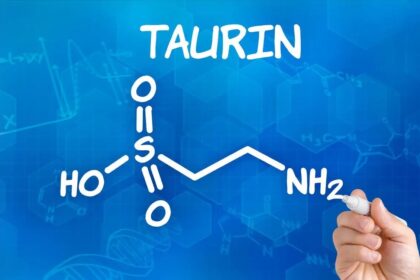
Iron is an essential trace element for the human body, but iron overload caused by long-term excessive intake (especially via supplements) disrupts the dynamic balance between bone formation and bone resorption through three pathways—oxidative stress, interference with mineral metabolism, and disruption of hormonal balance—ultimately leading to decreased BMD and increased risk of osteoporosis. Compared with traditional inorganic iron (e.g., ferrous sulfate), ferrous gluconate has a milder effect on bones due to the slow release of Fe²⁺; however, vigilance is still needed for long-term high-dose use.
(I) Core Mechanisms of BMD Reduction
1. Oxidative Stress Damages Osteoblasts
Excess iron ions (Fe²⁺) in the body easily generate hydroxyl radicals (・OH) through the Fenton reaction. These radicals directly damage the DNA and mitochondria of osteoblasts, inhibiting osteoblast proliferation and the synthesis of bone matrix (e.g., reduced secretion of type I collagen and osteocalcin). Studies have shown that in populations taking ferrous gluconate long-term (with daily elemental iron > 60 mg), the serum level of malondialdehyde (MDA)—a marker of oxidative damage—is 18%-25% higher than that in healthy individuals, while the level of bone-specific alkaline phosphatase (BALP)—a marker of osteoblast activity—is 12%-15% lower, indicating suppressed osteoblastic function.
2. Interference with Calcium-Phosphorus Metabolism and Vitamin D Activity
Iron overload competitively inhibits intestinal calcium absorption (iron and calcium share the divalent metal transporter 1 [DMT1] in the intestinal mucosa) and reduces the conversion efficiency of 25-hydroxyvitamin D to its active form (1,25-dihydroxyvitamin D) in the kidneys. Active vitamin D is a key hormone that promotes calcium absorption and bone mineralization; a decrease in its level leads to insufficient calcium deposition in the bone matrix and gradual reduction in BMD. Clinical data show that in populations taking ferrous gluconate continuously for more than 12 months without additional calcium and vitamin D supplementation, serum calcium concentration decreases by 5%-8% and 25-hydroxyvitamin D level drops by 10%-12% compared to baseline.
3. Exacerbation of Osteoclast Activity
Excess iron activates the NF-κB inflammatory pathway, promoting the expression of receptor activator of nuclear factor kappa-B ligand (RANKL) while inhibiting the secretion of osteoprotegerin (OPG)—a factor that suppresses osteoclasts. This leads to excessive osteoclast activation and accelerated bone resorption. Bone biopsies of individuals on long-term iron supplementation reveal that the number of osteoclasts in those with iron overload is 20%-30% higher than in healthy individuals, and the level of β-C-terminal telopeptide of type I collagen (β-CTX)—a marker of bone resorption—is 15%-20% higher, indicating a significant imbalance between bone resorption and bone formation.
(II) Characteristics of High-Risk Populations
Not all individuals on long-term ferrous gluconate use will experience BMD issues; the following groups face higher risks:
Elderly individuals (> 65 years old): Elderly people already have reduced osteoblast activity and accelerated bone loss. Oxidative stress caused by long-term iron supplementation further exacerbates bone metabolism imbalance, making their risk of BMD reduction 2-3 times higher than that of middle-aged and young adults.
Postmenopausal women: Decreased estrogen levels after menopause already increase the risk of osteoporosis due to enhanced bone resorption. Iron overload amplifies this effect, accelerating the rate of BMD loss in the lumbar spine and hip (an additional 1%-1.5% annual loss).
Populations not supplementing with calcium and vitamin D synchronously: Without additional calcium (daily intake < 800 mg) and vitamin D (daily intake < 400 IU) during long-term iron supplementation, intestinal calcium absorption disorders and insufficient bone mineralization become more prominent, significantly increasing the risk of BMD reduction.
II. Cardiovascular Risks of Long-Term Ferrous Gluconate Use: Mechanisms and Clinical Manifestations
The harm of long-term iron overload to the cardiovascular system mainly stems from oxidative stress-induced vascular endothelial damage, promotion of atherosclerosis, and interference with myocardial metabolism by iron ions, which may increase the risks of hypertension, atherosclerosis, and arrhythmia. Although ferrous gluconate has a lower cardiovascular risk than ferrous sulfate (as ferrous sulfate causes more rapid iron overload) due to its gradual iron release, potential risks still require vigilance during long-term excessive use.
(I) Core Mechanisms of Cardiovascular Risks
1. Vascular Endothelial Function Damage
Vascular endothelial cells act as a "protective barrier" for the cardiovascular system. ・OH generated by excess Fe²⁺ damages the integrity of endothelial cells, inhibits the synthesis of nitric oxide (NO)—a key substance for maintaining vascular relaxation and inhibiting platelet aggregation—and promotes the secretion of endothelin-1 (ET-1), a potent vasoconstrictor. This leads to vascular diastolic dysfunction and increased hypertension risk. Clinical studies have found that in populations taking ferrous gluconate continuously for more than 18 months (daily elemental iron > 50 mg), flow-mediated dilation (FMD) of the brachial artery—a measure of endothelial function—decreases by 8%-10% compared to baseline, and systolic blood pressure increases by an average of 5-8 mmHg.
2. Promotion of Atherosclerotic Plaque Formation
Iron overload accelerates atherosclerosis through two pathways:
Oxidation of low-density lipoprotein (ox-LDL): Fe²⁺ oxidizes LDL in the blood to ox-LDL, which is easily phagocytosed by macrophages to form foam cells. These cells accumulate in the vascular wall, forming atherosclerotic plaques.
Exacerbation of inflammatory responses: Iron ions activate macrophages to release inflammatory factors such as tumor necrosis factor-α (TNF-α) and interleukin-6 (IL-6), promoting plaque progression and instability.
Lipid monitoring in individuals on long-term iron supplementation shows that ox-LDL levels in those with iron overload are 20%-25% higher than in healthy individuals, and carotid intima-media thickness (IMT)—an early marker of atherosclerosis—increases by 0.1-0.2 mm.
3. Myocardial Cell Damage and Arrhythmia
Myocardial cells are highly sensitive to iron overload. Excess iron ions accumulate in myocardial mitochondria, disrupting mitochondrial function and causing myocardial energy metabolism disorders. Meanwhile, oxidative stress induced by iron ions damages the myocardial cell membrane, interfering with ion balance (e.g., calcium and potassium ion disorders) and increasing the risk of arrhythmias such as ventricular premature beats and atrial fibrillation. Animal experiments confirm that mice with long-term iron overload have 30%-40% higher myocardial iron content and 15%-20% higher myocardial cell apoptosis rate than normal mice, and electrocardiograms show a 2-3 fold increase in ventricular premature beat incidence.
(II) Characteristics of High-Risk Populations
The following groups face higher cardiovascular risks when taking ferrous gluconate long-term:
Individuals with pre-existing cardiovascular diseases: Patients with hypertension, coronary heart disease, or diabetes face aggravated pathological damage from iron overload, which accelerates disease progression (e.g., increased risk of plaque instability in coronary heart disease patients).
Patients with chronic kidney disease (CKD): CKD patients already have iron metabolism disorders (insufficient erythropoietin leads to impaired iron utilization, often requiring iron supplementation to correct anemia). Long-term iron supplementation easily causes iron overload, making their cardiovascular risk 3-4 times higher than that of the general population.
Long-term alcohol consumers: Alcohol enhances iron absorption and storage in the liver and exacerbates oxidative stress induced by iron ions. Combined use with ferrous gluconate significantly increases cardiovascular risks (e.g., alcoholic cardiomyopathy combined with iron-induced myocardial damage).
III. Safety Monitoring Protocol for Long-Term Ferrous Gluconate Use
To avoid BMD reduction and cardiovascular risks caused by long-term ferrous gluconate use, a comprehensive management protocol combining "regular indicator monitoring, dynamic dosage adjustment, and lifestyle intervention" is required. The core monitoring indicators and frequencies are as follows:
(I) BMD-Related Monitoring
1. Basic Indicator Monitoring (Every 6 Months)
Serum iron metabolism indicators: Including serum iron (SI), serum ferritin (SF), and transferrin saturation (TSAT). Normal ranges: SI = 10-30 μmol/L; SF = 30-400 μg/L (males), 15-200 μg/L (females); TSAT = 20%-45%. If SF > 400 μg/L (males) or > 200 μg/L (females), or TSAT > 45%, iron overload is indicated, and the iron supplementation dosage must be reduced immediately (e.g., from 60 mg elemental iron daily to 30 mg).
Bone metabolism markers: Including bone formation markers (BALP, normal range = 40-150 U/L; osteocalcin [OC], normal range = 5-20 ng/mL) and bone resorption marker (β-CTX, normal range = 0.2-1.0 ng/mL). If BALP/OC decreases or β-CTX increases, bone metabolism imbalance is indicated, requiring supplementation with calcium (800-1000 mg daily) and vitamin D (400-800 IU daily).
2. BMD Imaging Monitoring (Every Year)
Dual-energy X-ray absorptiometry (DXA): Used to measure BMD of the lumbar spine (L1-L4) and hip (femoral neck), and calculate the T-score. Normal: T-score ≥ -1.0; osteopenia: -2.5 < T-score < -1.0; osteoporosis: T-score ≤ -2.5. If osteopenia occurs, the iron supplementation protocol must be adjusted under medical guidance, and anti-resorptive interventions (e.g., bisphosphonate supplementation, strictly following medical advice) should be added.
(II) Cardiovascular-Related Monitoring
1. Basic Indicator Monitoring (Every 3-6 Months)
Blood pressure and heart rate: Measure blood pressure at a fixed time weekly (10 minutes after morning fasting rest). Normal blood pressure should be controlled below 130/80 mmHg. If blood pressure persists above 130/80 mmHg, investigate whether iron overload is a contributing factor and adjust the iron supplementation dosage if necessary.
Lipid and oxidative stress indicators: Including total cholesterol (TC), low-density lipoprotein cholesterol (LDL-C), high-density lipoprotein cholesterol (HDL-C), ox-LDL, and serum MDA. If LDL-C > 3.4 mmol/L, ox-LDL > 60 U/L, or MDA > 5.0 nmol/mL, the risk of atherosclerosis is elevated. Adjust the diet (reduce high-fat, high-sugar foods) under medical guidance, and add lipid-lowering drugs (e.g., statins, following medical advice) if necessary.
Vascular endothelial function indicator: Measure FMD of the brachial artery via ultrasound. Normal FMD ≥ 7%. If FMD < 7%, vascular endothelial dysfunction is indicated; reduce the iron supplementation dosage and supplement with antioxidants (e.g., 100-200 mg vitamin C daily, 10-15 mg vitamin E daily).
2. Cardiovascular Imaging and Function Monitoring (Every Year)
Carotid ultrasound: Measures carotid IMT. Normal IMT < 1.0 mm. If IMT ≥ 1.0 mm, early atherosclerosis is indicated; further coronary CTA is required to screen for plaques.
Additional tests for patients with pre-existing cardiovascular diseases: Electrocardiogram (ECG) and echocardiography to monitor myocardial structure and function, preventing iron overload-induced myocardial damage.
IV. Safety Management Strategies for Long-Term Use
In addition to regular monitoring, risks can be further reduced through "precise dosage control, combined intervention, and lifestyle adjustment." Specific strategies are as follows:
(I) Precisely Control Iron Supplementation Dosage to Avoid Iron Overload
Follow the "supplement only what is deficient" principle: Regularly test SF and TSAT. Once anemia is corrected (Hb returns to normal: > 130 g/L for males, > 120 g/L for females) and SF reaches the normal range, reduce the "therapeutic dosage" to a "maintenance dosage" (e.g., 30-40 mg elemental iron daily), avoiding long-term use of therapeutic dosages (> 60 mg elemental iron daily).
Avoid combined use with other iron supplements: During long-term ferrous gluconate use, do not take other iron supplements (e.g., ferrous sulfate, ferrous fumarate) simultaneously. Also, reduce excessive intake of iron-rich foods (e.g., animal liver, animal blood) to 1-2 times weekly (50-100 g per serving) to prevent overlapping iron intake.
(II) Combined Nutrient Supplementation to Counteract Iron Overload Harm
Synchronously supplement calcium and vitamin D: Take 800-1000 mg calcium daily (in two doses, with a 2-hour interval from iron supplements to avoid absorption competition) and 400-800 IU vitamin D daily (promotes calcium absorption and bone mineralization) to counteract iron-induced interference with bone metabolism.
Supplement antioxidants: Take 100-200 mg vitamin C daily (taken with iron supplements to improve iron absorption while counteracting oxidative stress) and 10-15 mg vitamin E daily to reduce iron-induced vascular endothelial damage and atherosclerosis risk.
(III) Adjust Lifestyle to Strengthen Safety Assurance
Diet management: Reduce intake of high-fat, high-sugar, and high-salt foods; increase intake of fiber-rich foods (e.g., whole grains, vegetables); avoid alcohol (alcohol exacerbates iron overload and liver damage).
Exercise intervention: Perform moderate-intensity exercise 3-5 times weekly (e.g., brisk walking, swimming, yoga, 30-45 minutes per session). Exercise promotes bone formation (stimulates osteoblast activity) and vascular health (improves endothelial function), but avoid excessive exercise (e.g., high-intensity interval training) to prevent increased physical stress.
The core of long-term ferrous gluconate safety lies in avoiding iron overload—oxidative stress and inflammatory responses caused by excess iron are key factors leading to decreased BMD and elevated cardiovascular risks. Early risk detection can be achieved through "regular monitoring of iron metabolism, BMD, and cardiovascular indicators," while harms can be effectively avoided through "precise dosage control, combined calcium and vitamin D supplementation, and lifestyle adjustment."
It is important to note that risk levels vary significantly among populations (e.g., elderly individuals, postmenopausal women, and patients with cardiovascular diseases face higher risks), requiring personalized monitoring and intervention protocols. In general, with standardized use and scientific monitoring under medical guidance, the long-term safety of ferrous gluconate is controllable. It can correct iron deficiency while minimizing BMD and cardiovascular risks, providing safety assurance for populations requiring long-term iron supplementation.