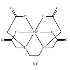
As a clinically commonly used organic iron supplement, the core of ferrous gluconate’s regulatory effect on the immune system lies in providing the body with highly absorbable iron. Iron is an "essential trace element" for maintaining the normal operation of the immune system— the proliferation, differentiation, and effector functions of immune cells all rely on iron as an enzyme cofactor, signaling molecule, or metabolic substrate. Iron deficiency (a common condition in patients with iron-deficiency anemia) directly leads to impaired immune function. Supplementing ferrous gluconate can correct iron deficiency, provide a material basis for the recovery and activation of immune cell functions, and thereby achieve positive regulation of the immune system.
I. Iron: The "Core Support Factor" for Immune Cell Functions
The efficient operation of the immune system essentially involves immune cells (e.g., phagocytes, T cells, B cells, natural killer cells) collaborating to complete the process of "recognizing pathogens, activating responses, and eliminating foreign substances." Every link in this process is inseparable from iron. Iron deficiency disrupts the metabolic balance of immune cells, leading to their "inactivation" or "low efficiency," which is specifically manifested as: decreased proliferation rate of immune cells, weakened bactericidal ability, and reduced antibody production. As an organic iron with good water solubility and high bioavailability (compared with inorganic iron such as ferrous sulfate, it causes less gastrointestinal irritation and its absorption is not significantly affected by dietary factors like phytic acid), ferrous gluconate can be more efficiently absorbed by the intestines and transported to immune organs (e.g., spleen, lymph nodes, thymus), supplementing "functional iron" for immune cells and repairing immune functions damaged by iron deficiency.
II. Regulatory Effects of Ferrous Gluconate on Key Immune Cell Functions
1. On Phagocytes (Neutrophils, Macrophages): Strengthening the "Pathogen Clearance Line of Defense"
Phagocytes are the "first line of defense" of the immune system, and their core function is to phagocytose and kill invading pathogens such as bacteria and fungi. Both key links of this process rely on iron:
Energy and "Weapon Supply" for Phagocytosis and Bactericidal Activity: After phagocytosing pathogens, phagocytes generate a large amount of reactive oxygen species (ROS, such as superoxide anions and hydrogen peroxide) through "respiratory burst" to kill pathogens. This process requires catalysis by nicotinamide adenine dinucleotide phosphate (NADPH) oxidase, and iron is the core cofactor of this enzyme. In iron deficiency, the activity of NADPH oxidase decreases significantly, weakening the "respiratory burst" capacity of phagocytes, which fail to effectively kill phagocytosed pathogens, increasing susceptibility to infection. After supplementing ferrous gluconate, iron can activate NADPH oxidase and restore the bactericidal efficiency of phagocytes.
"Catalyst" for Enzymatic Reactions: The activity of bactericidal enzymes in macrophages, such as myeloperoxidase (MPO) and lysozyme, also depends on iron. For example, MPO can use ROS to generate hypochlorous acid (HClO) with strong bactericidal effects, and sufficient iron maintains the conformational stability and activity of MPO, enhancing macrophages’ ability to clear intracellular pathogens (e.g., Mycobacterium tuberculosis). In addition, macrophages participate in the "iron sequestration" defense mechanism—they "sequester" free iron in the body by synthesizing iron-binding proteins (e.g., lactoferrin), preventing bacteria from obtaining iron (bacterial growth relies on iron). In this process, the iron metabolic balance of macrophages themselves also requires exogenous iron (such as that provided by ferrous gluconate) to maintain, avoiding the failure of the defense mechanism due to their own iron deficiency.
2. On T Lymphocytes: Maintaining the "Regulatory Hub of Immune Responses"
T cells are the "regulatory hub" of the immune system, responsible for recognizing pathogen antigens, activating other immune cells (e.g., B cells, phagocytes), and directly killing pathogen-infected target cells. Their activation, proliferation, and differentiation rely on iron throughout the process:
"Signal Switch" for T Cell Activation: After T cells recognize antigens through T cell receptors (TCR), they need to achieve activation through intracellular signaling pathways (e.g., PI3K-Akt, MAPK pathways). The activity of key kinases in these pathways (e.g., Akt kinase) depends on iron as a cofactor. In iron deficiency, T cell signaling pathway transduction is blocked, and T cells cannot switch from a "quiescent state" to an "activated state," leading to delayed initiation of immune responses. After supplementing ferrous gluconate, iron can promote the activation of T cell signaling pathways and accelerate T cells’ recognition and response to pathogens.
"Metabolic Fuel" for T Cell Proliferation and Differentiation: After activation, T cells need to proliferate in large quantities to form sufficient effector T cells (e.g., cytotoxic T cells, helper T cells). This process relies on DNA synthesis and energy metabolism—iron is an essential component of DNA polymerase (involved in DNA replication) and mitochondrial respiratory chain enzymes (e.g., cytochrome c oxidase, responsible for ATP production). Iron deficiency reduces the proliferation rate of T cells and causes differentiation imbalance: helper T cells (Th cells) shift toward Th2-type cells (biased toward humoral immunity), while the proportion of Th1-type cells (biased toward cellular immunity, responsible for clearing intracellular pathogens) decreases, weakening the body’s resistance to viruses and intracellular bacterial infections. By supplementing iron, ferrous gluconate can restore the normal proliferation rhythm of T cells, regulate Th1/Th2 balance, and enhance the synergy between cellular and humoral immunity.
"Killing Weapon" of Cytotoxic T Cells: Cytotoxic T cells kill infected cells by releasing perforin (forming membrane pores) and granzyme (inducing target cell apoptosis), and the synthesis and activity of granzyme depend on iron. In iron deficiency, granzyme secretion decreases, significantly reducing the killing efficiency of T cells; iron supplementation can promote granzyme synthesis and enhance their ability to clear target cells.
3. On B Lymphocytes: Promoting the "Effector Output of Humoral Immunity"
The core function of B cells is to differentiate into plasma cells and produce specific antibodies (the core effector molecules of humoral immunity), and this process also requires iron support:
Proliferation and Differentiation of B Cells: After being stimulated by antigens, B cells need to undergo proliferation and differentiation into plasma cells. DNA replication and protein synthesis (antibodies are essentially proteins) during this process rely on iron—iron participates in ribosome assembly (ribosomes are the sites of protein synthesis) and is a cofactor of amino acid synthetases. Iron deficiency hinders B cell proliferation, reduces the number of plasma cells, and directly leads to decreased production of antibodies (e.g., IgG, IgM). As a result, the body cannot neutralize pathogens (e.g., viruses) through antibodies or mark bacteria for clearance by phagocytes.
Antibody Affinity Maturation: The "quality" (affinity) of antibodies depends on gene mutation and selection of B cells during differentiation, which requires cells to maintain active metabolism. By maintaining mitochondrial function (providing energy) and enzyme activity, iron ensures the metabolic activity of B cells, indirectly promoting antibody affinity maturation and enabling the body to produce antibodies that bind to pathogens more efficiently.
4. On Natural Killer (NK) Cells: Enhancing the "Rapid Defense of Innate Immunity"
NK cells are key cells of innate immunity that can directly kill tumor cells or virus-infected cells without prior antigen stimulation. Their cytotoxicity relies on iron as a "functional cofactor":
The cytotoxic effect of NK cells depends on the perforin-granzyme system and the Fas-FasL pathway, both of which require iron to activate related enzymes (e.g., caspases). In iron deficiency, the ability of NK cells to release perforin and granzyme decreases, significantly reducing their killing rate of target cells; supplementing ferrous gluconate allows iron to restore the cytotoxicity of NK cells and enhance the body’s ability to quickly clear abnormal cells.
In addition, the activation of NK cells is regulated by cytokines (e.g., IL-2, IFN-γ), and iron can promote T cells to secrete cytokines such as IL-2, indirectly providing "activation signals" for NK cells and further enhancing their defense function.
III. The Role of Iron in Immune Signaling Pathways and Immune Response Regulation
In addition to directly supporting immune cell functions, iron also affects the intensity and direction of overall immune responses by regulating immune-related signaling pathways:
Regulatory Role of Iron Regulatory Proteins (IRPs): Intracellular iron levels are sensed by IRPs. In iron deficiency, IRPs bind to the mRNA of immune-related genes (e.g., genes encoding lactoferrin and transferrin receptors) to regulate their expression—for example, promoting transferrin receptor synthesis (enhancing iron absorption) while inhibiting ferritin synthesis (reducing iron storage) to prioritize meeting the iron needs of immune cells. After supplementing ferrous gluconate, iron levels increase, inhibiting IRP activity and instead promoting ferritin synthesis (storing excess iron), avoiding iron-induced damage to immune cells and maintaining the balance between iron metabolism and immune function.
Interaction Between Iron and Cytokines: Cytokines (e.g., IL-2, IFN-γ, TNF-α) are "signal messengers" between immune cells, and their synthesis and secretion depend on iron. For example, IFN-γ secreted by T cells can promote macrophages to express transferrin receptors, enhancing iron uptake and thereby improving macrophages’ bactericidal ability; in turn, sufficient iron can promote T cells to secrete IFN-γ, forming a positive regulatory cycle of "iron-cytokine-immune cells." By supplementing iron, ferrous gluconate can strengthen this cycle and enhance the synergy of immune responses.
IV. "Bidirectionality": Appropriate Supplementation Is the Key to Exerting Immune Regulatory Effects
Iron’s regulation of the immune system is "bidirectional," and the immune regulatory effect of ferrous gluconate depends on "appropriate supplementation." Both excess and deficiency can impair immune function:
Harm of Iron Deficiency: As mentioned earlier, iron deficiency leads to a comprehensive decline in the proliferation, differentiation, and effector functions of immune cells, manifested as weakened resistance to infection (susceptibility to recurrent colds and persistent infections) and insufficient antibody response after vaccination (reduced vaccine protection).
Risks of Excess Iron: Blind excessive supplementation of ferrous gluconate (exceeding the daily recommended amount—adults require approximately 12–20 mg of iron per day) leads to accumulation of unused iron in immune cells, which instead has negative effects: On one hand, excess iron generates a large number of hydroxyl radicals (・OH) through the Fenton reaction, triggering oxidative stress, damaging the cell membranes, proteins, and DNA of immune cells, and inhibiting their functions. On the other hand, the growth of pathogens such as bacteria and viruses also requires iron (e.g., bacteria absorb iron through siderophores), and excess iron provides "nutrition" for pathogens, potentially promoting infection spread (e.g., Mycobacterium tuberculosis multiplies more easily in iron-sufficient environments). In addition, long-term iron overload may induce chronic inflammation, further disrupting immune homeostasis.
V. Summary and Application Precautions
The regulatory effect of ferrous gluconate on the immune system essentially involves efficiently supplementing iron to repair immune cell function defects caused by iron deficiency, strengthening the proliferation, differentiation, and effector functions of key immune cells (phagocytes, T cells, B cells, NK cells), and maintaining the balance and efficiency of immune responses by regulating iron-related signaling pathways and cytokine networks. Its advantage lies in being an organic iron with high bioavailability and good tolerance, which can accurately provide "functional iron" for immune cells.
However, it should be clarified that the immune regulatory effect of ferrous gluconate is mainly targeted at iron-deficient populations (e.g., patients with iron-deficiency anemia, individuals with iron deficiency due to chronic blood loss, and those with insufficient dietary iron intake). For people with sufficient iron nutrition, additional supplementation cannot enhance immune function; instead, it may trigger risks due to iron excess. Therefore, its use must follow the principle of "supplementation on demand": When used as an auxiliary means of immune regulation, it is necessary to first confirm the presence of iron deficiency through tests (e.g., serum ferritin, hemoglobin), then determine the dosage under the guidance of doctors or dietitians, and avoid blind supplementation. Only in this way can we truly achieve scientific regulation of immune function "with iron as the medium."